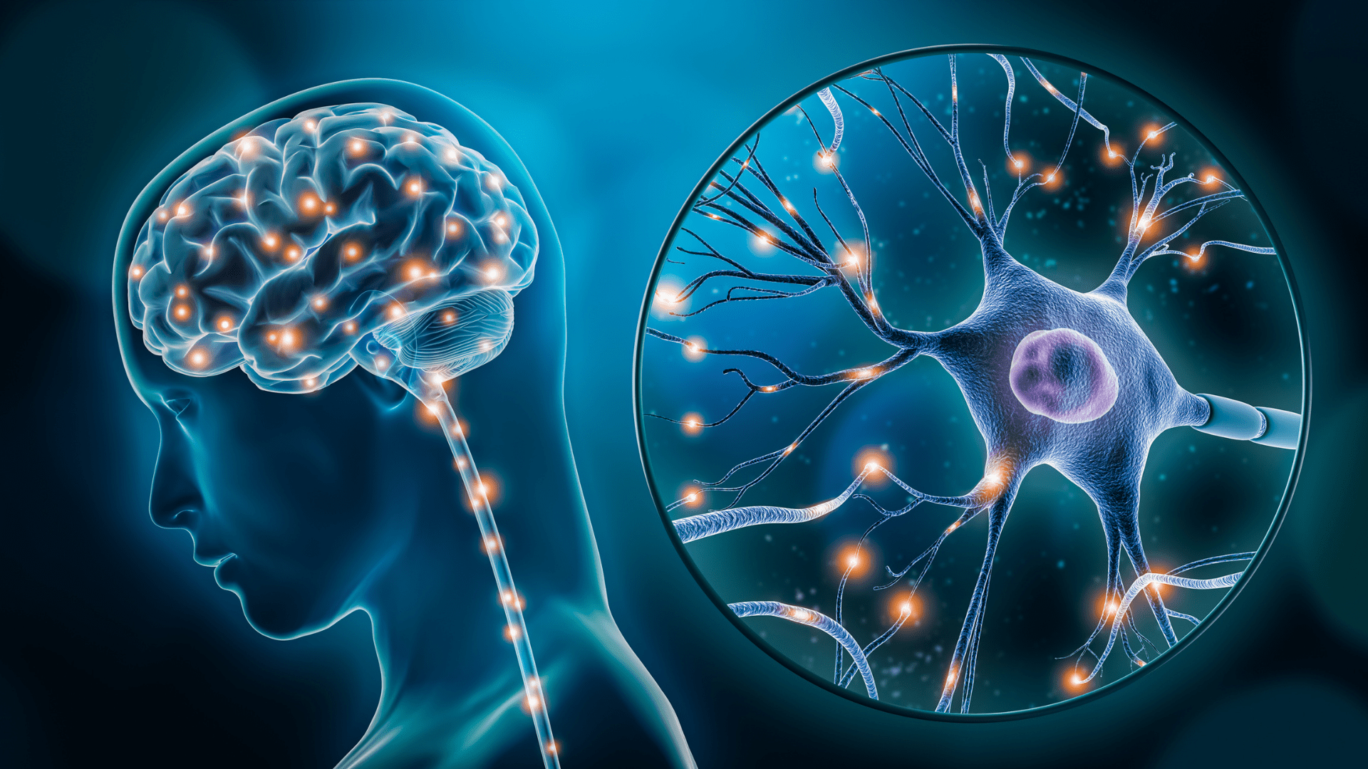
Scientists are in the process of finding out more about the brain and its tendencies. specifically when brain cells shut off. They’re looking at the brain’s “off switches.” This new technology tracks when brain cells switch off, usually after a burst of activity, known as inhibition. Until now, it’s been a mystery how long neurons stay active and when they turn off.
Scientists at Scripps Research came up with a new technique to study why the brain’s “off switches” go out of control in normal behavior, in diseases and disorders.
Inhibition

It is agreed that inhibition of neurons is how the brain regulates activities but finding a way to track it proved to be difficult. Until now, only a few have been able to look at it in a trackable way. Li Ye and John Yates are both professors at Scripps Research. Together, they are studying how brain cells change when they’re actively firing and comparing them to when they are done firing. When a brain cell fires, it is passing a message through an electrical charge.
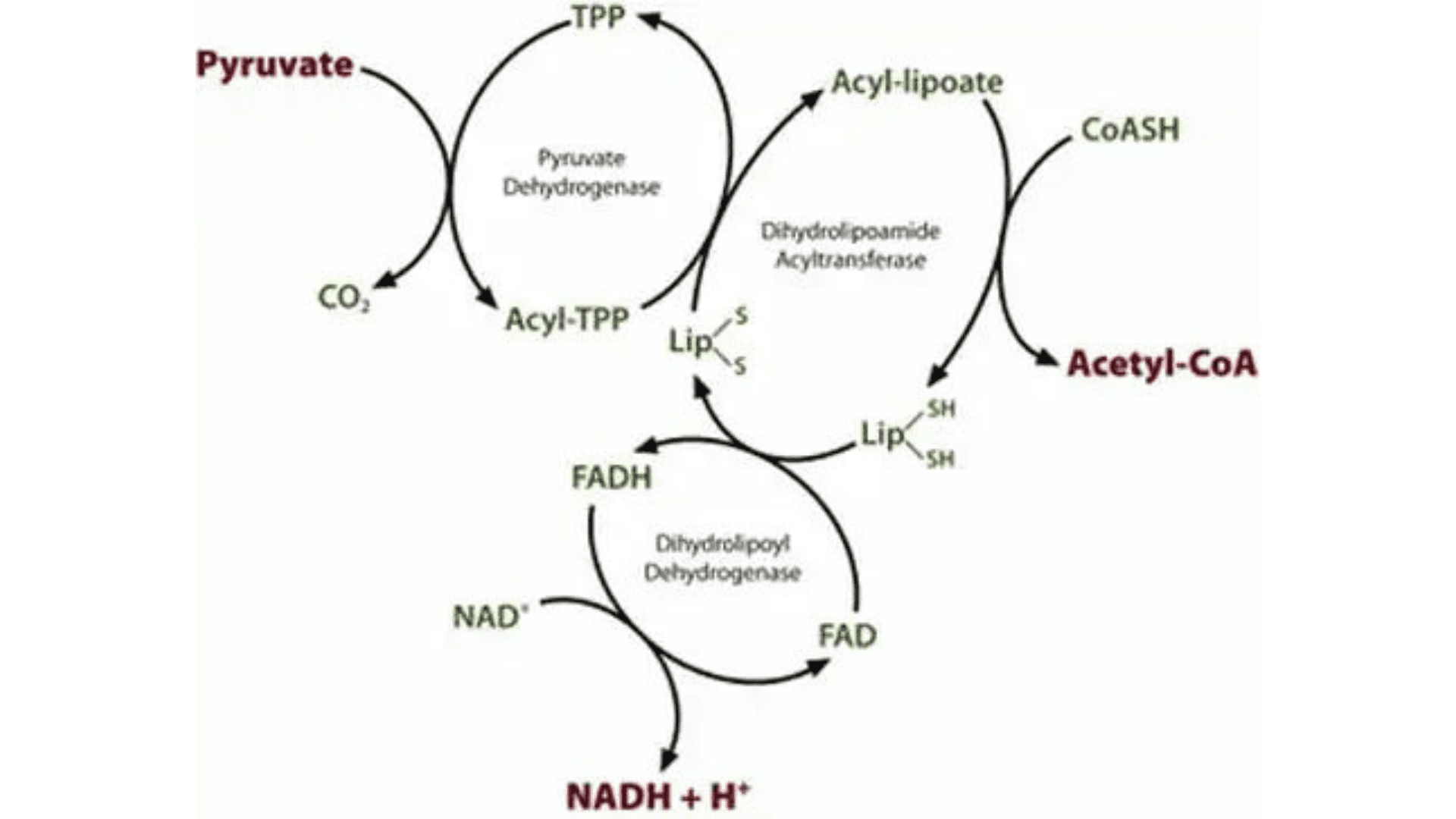
Proteins are important in this process. To measure the levels and characteristics of the modifications of these proteins, scientists use optogenetics. This allows them to encode information from selected cells. Through this process, they identified a protein called pyruvate dehydrogenase (PDH). PDH rapidly changes immediately after brain cells are inhibited.
PDH Protein

Explore Tomorrow's World from your inbox
Get the latest innovations shaping tomorrow’s world delivered to your inbox!
I understand that by providing my email address, I agree to receive emails from Tomorrow's World Today. I understand that I may opt out of receiving such communications at any time.
Ye said, “When neurons are firing, you need a lot of energy, and this PDH protein is involved in producing that energy” but “the brain really wants to conserve energy, so when a cell is done firing, we found that the brain rapidly shuts off the PDH protein. This happened much faster than anything else we saw in gene expression.”
Researchers found out that the cells add molecular tags to the protein to shut off the PDH. These are called phosphates. The scientists also found antibodies that only recognized the inactive, form of PDH with the phosphates (pPDH). To test if the levels of pPDH could be used to act on behalf of brain cell inhibition, Ye and his team used the antibodies to measure pPDH in mice that were under anesthesia. When they looked at the results, nearly the entire brain lit up with high levels of pPDH. This shows most of the brain is inactive under anesthesia.
Asking questions
A lot of questions pop up because of the discovery. Ye asked, “If the brain can’t turn cells off, or if they’re turned off faster or slower than usual, what happens? How does the inhibition of neurons play a role in different diseases?”
Their new method could help find out what goes wrong in different brain conditions like depression, post-traumatic stress disorder, and Alzheimer’s disease.


