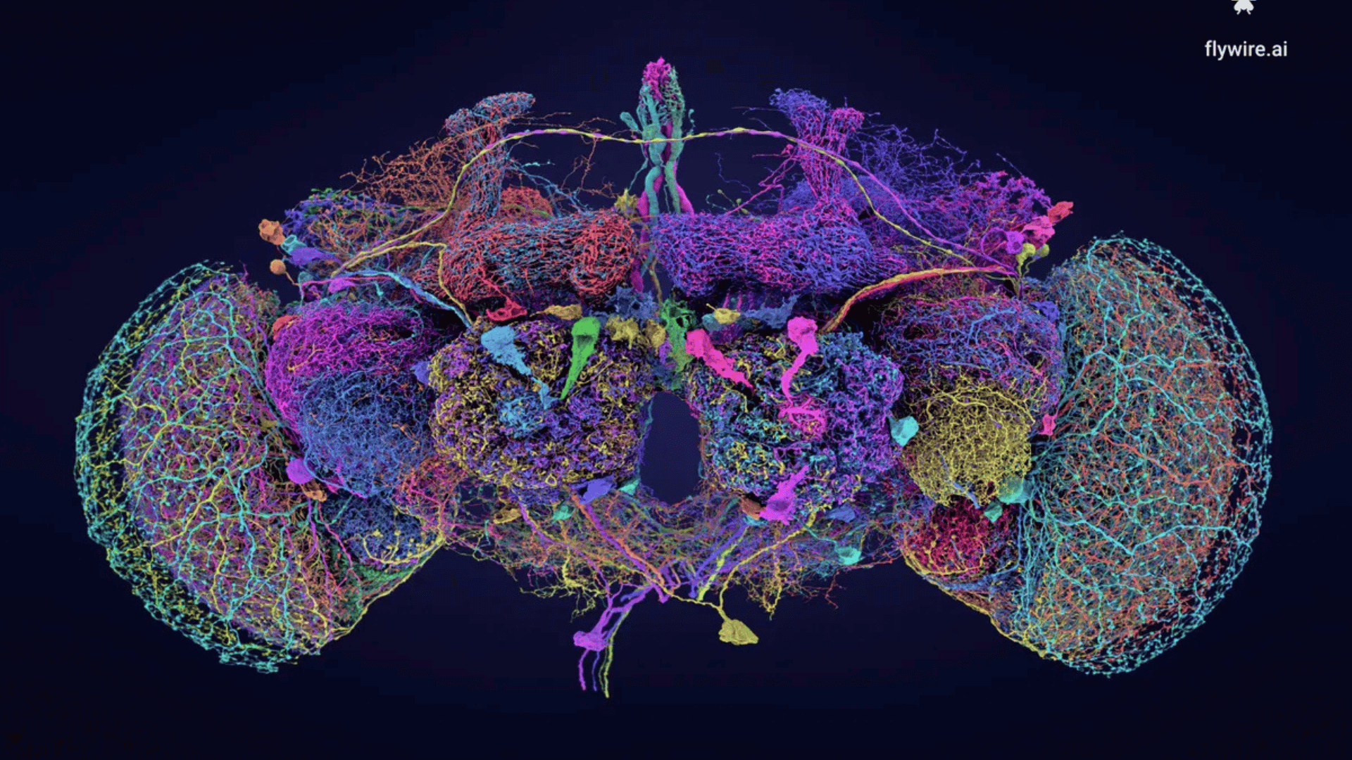
An international research consortium has unveiled the first complete map of a fruit fly brain, progressing us one step closer to mapping the human brain.

Maps that display every neuron in the brain and the connections between them are called connectomes. The first organism to be mapped in this way was the worm Caenorhabditis elegans, but it doesn’t have a brain but rather a “nerve ring” so it’s not a good representation of the complexity of the human brain.
“Flies can do all kinds of complicated things like walk, fly, navigate, and the males sing to the females,” explained Dr Gregory Jefferis of the University of Cambridge, one of the co-leaders of the new research, in a statement.
Researchers chose the Drosophila melanogaster fruit fly because of its complex brain, which has 139,255 neurons and 50 million synapses. Even this quantity is still small compared to the human brain, which is thought to contain closer to 80 billion neurons.
“If we want to understand how the brain works, we need a mechanistic understanding of how all the neurons fit together and let you think,” continued Jefferis. “For most brains, we have no idea how these networks function.”
The FlyWire Consortium was launched to leverage cutting-edge technology and expertise from labs around the world to create a full connectome of the fruit fly brain.
The map began with 21 million images of an adult female fruit fly brain. Next, the team used artificial intelligence (AI) to align the images and generate 3D reconstructions of the individual neurons. Hundreds of researchers and citizen scientists worldwide were enlisted to help proofread the map.
Additional existing data was used to identify and categorize the 50 million synapses. Finally, Jefferis, University of Vermont’s Davi Bock, and other colleagues annotated the different types of neurons that can be found within the brain.
Through the 3D animations, we can explore the fly’s auditory neurons or the cells the female uses to detect the songs of potential mates. We can also view the fly’s internal compass or EPG ring and CT1 neuron.
Though this is certainly a step toward a future where we could develop a similar type of map for humans, that’s still some time away as the human brain is vastly more complex.
“The fly brain is a milestone on our way to reconstructing a wiring diagram of a whole mouse brain,” stated Dr Sebastian Seung of Princeton University, one of the research co-leads.


